|
||
|
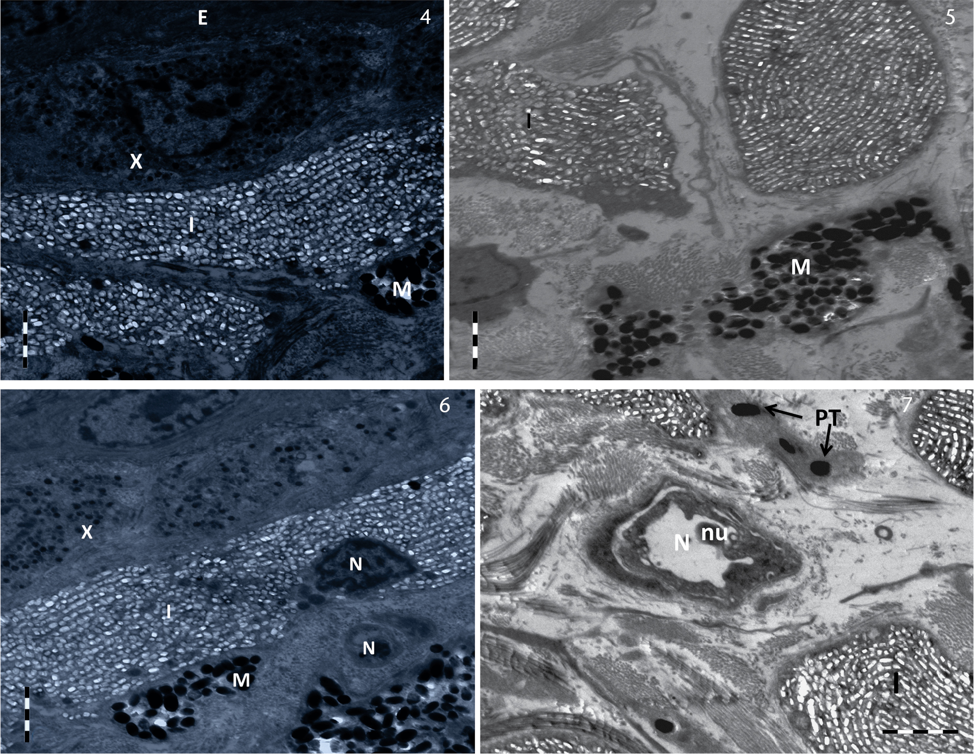
(4-5) Ultrastructural features of chromatophores of dorsal skin in A. orientalis. The vertical combination of dermal chromatophores is xanthophores at the top, iridophores in the middle, and melanophores at the bottom. (6-7) Electron photomicrograph showing the combination of dermal chromatophores in the skin of A. orientalis. (E) Epidermal layer, (I) iridophores, (M) melanophores, (nu) nucleolus, (N) nucleus, (PT) pterinosomes, (X) xanthophores. Scale bar: 2 μm. |