|
||
|
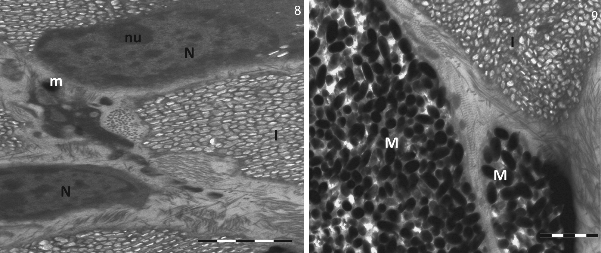
Electron photomicrograph showing different stages of melanosomes. (I) lridophores, (M) melanophores, (m) mitochondria, (N) nucleus, (nu) nucleolus. Scale bar: 2 μm. |
|
||||||||
| Part of: Paray BA, Al-Sadoon MK (2017) Ultrastructure of the dermal chromatophores in the Fringe-toed lizard, Acanthodactylus orientalis. Zoologia 34: 1-7. https://doi.org/10.3897/zoologia.34.e11923 |