|
||
|
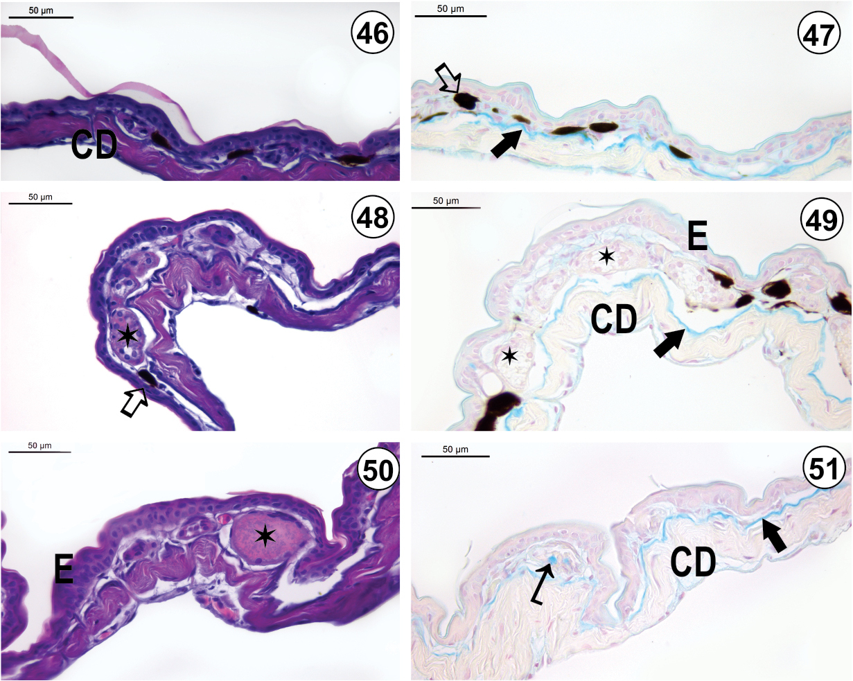
Light micrograph of the integument of O. v-signata: (46) Dorsal region (HE-staining); (47) Dorsal region (AB-method); (48) Ventrolateral region (HE-staining); (49) Ventrolateral region (AB-method); (50) Ventral region (HE-staining); (51) Ventral region (AB-method). Melanophores (_) occur in the spongious dermis that is poorly developed in the dorsal region. The EK-layer (Æ) is a continuous layer in all integument regions. Apocrine glands (¬) with heterogeneous content occur in both ventrolateral and ventral regions. Serous glands are visualized in both dorsal and ventrolateral integument. Mixed glands () are observed in the ventral region. E = epidermis; CD = compact dermis. |