|
||
|
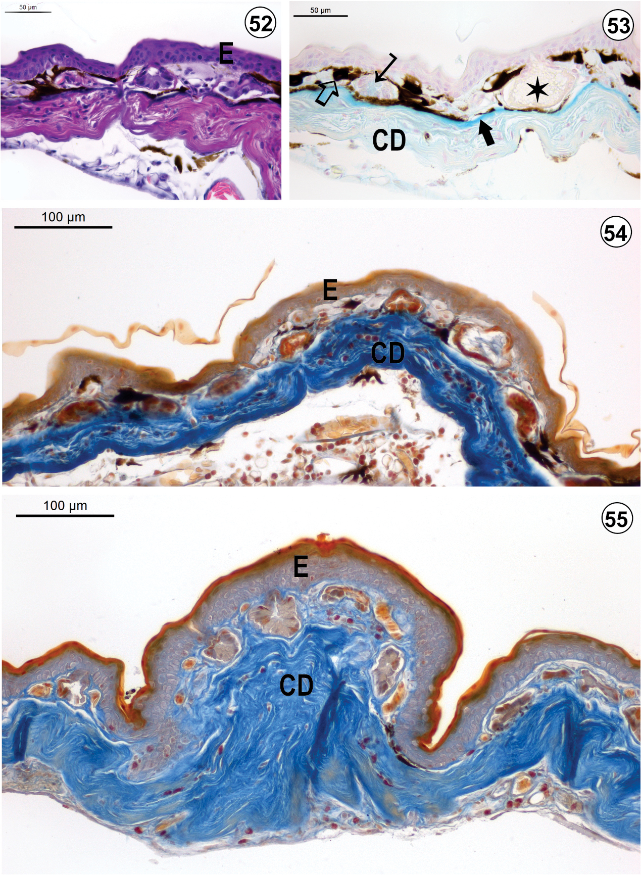
Light micrograph of the integument of S. x-signatus: (52) Dorsal region (HE-staining); (53) Dorsal region (AB-method); (54) Ventrolateral region (Mallory´s trichrome staining); (55) Ventral region (Mallory´s trichrome staining). The epidermis (E) is slightly ticker when compared to those of other hylids, as in the compact dermis (CD). Melanophores (_) are visulized in both dorsal and ventrolateral integument just beneath the epidermis. Serous glands are present in all regions; however, some of them show slightly alcianophilic content (→) in both ventrolateral and ventral regions. The apocrine glands (¬) with granular content occur in both dorsal and ventrolateral regions. The EK-layer (Æ) is visualized in both dorsal and ventrolateral integument but is absent in the ventral integument. |