|
||
|
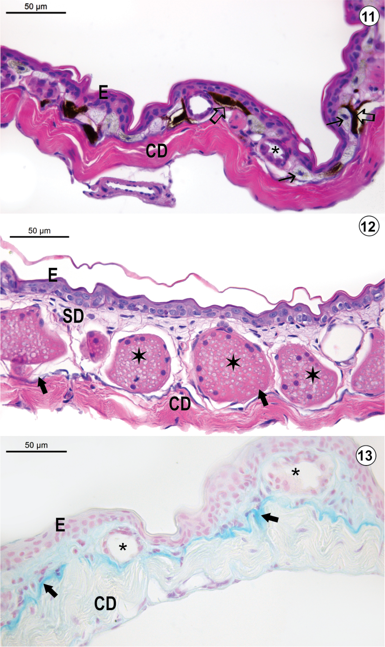
Light micrograph of the integument of O. albicans: (11) Dorsal region (HE-staining); (12) Ventrolateral region (HE-staining); (13) Ventral region (AB-method). In all integument regions, the epidermis (E) rests on the dermis, which is subdivided into the spongious dermis (SD) and the compact dermis (CD). Iridophores (→) occur in the dorsal region; however, they are absent in both ventrolateral and ventral regions. Melanophores (_) in the spongious dermis. Both serous (Ø) and apocrine glands (¬) occur in the spongious dermis. No glandular cell reacts to AB-method, suggesting that secretory units is made up of serous cells. The EK-layer (Æ) exhibits its typical basophilic staining. Note clusters of apocrine glands (¬) with heterogeneous content in the ventrolateral integument. The EK-layer () exhibits typical alcianophilic reaction of its glycoconjugate content. Large blood vessels occur in the hypodermis. |