|
||
|
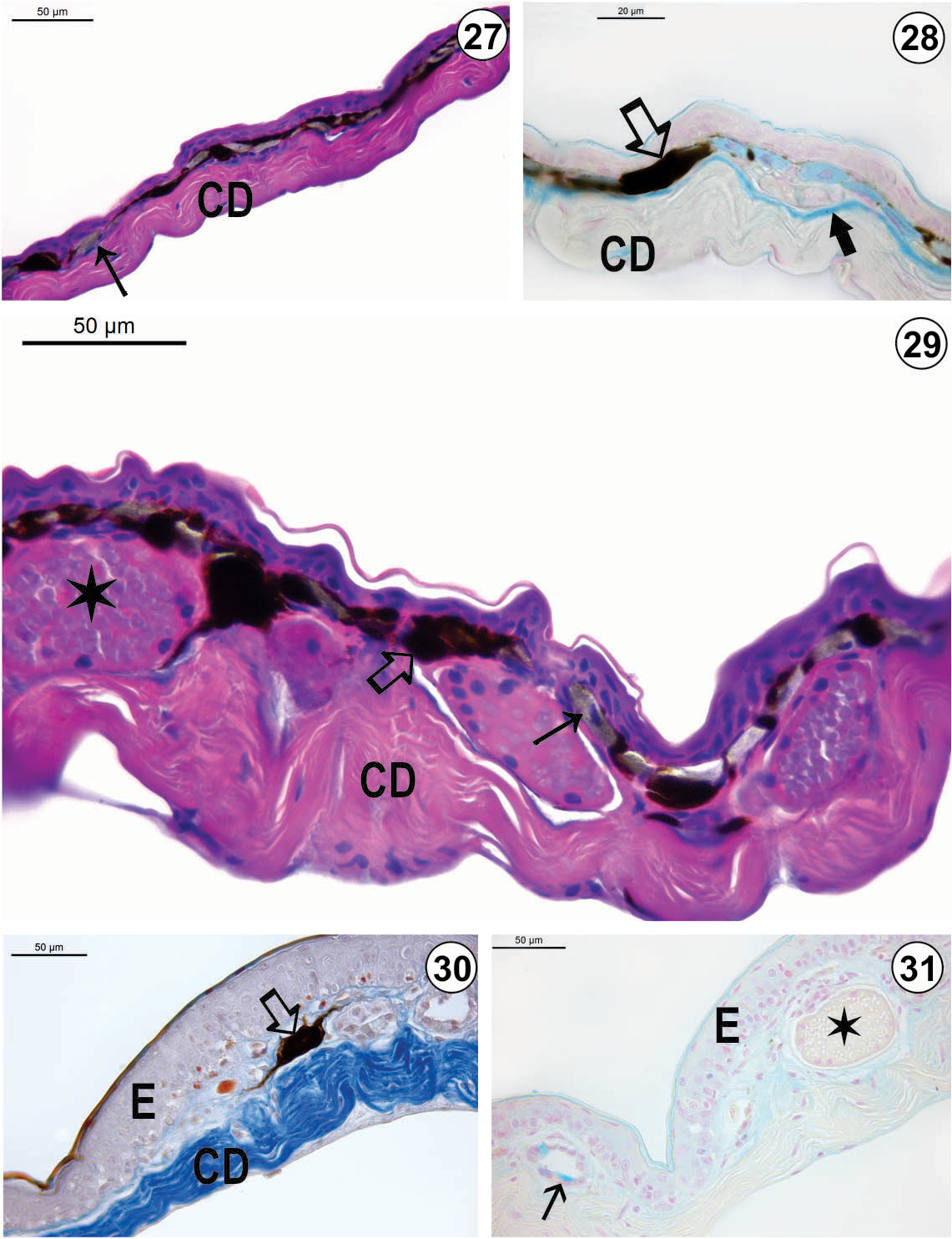
Light micrograph of the integument of O. humilis: (27) Dorsal region (HE-staining); (28) Dorsal region (AB-method); (29) Ventrolateral region (HE-staining) (30) Ventral region (Mallory´s trichrome staining); (31) Ventral region (AB-method). In the dorsal region, the spongious dermis is poorly developed. Melanophores (_) are visualized in all integument regions; however, iridophores (→) are visualized only in both dorsal and ventrolateral integument. Both pigment cells are located just beneath the epidermis. Alcianophilic reaction is observed in cytoplasm of iridophores as well as in the EK-layer (Æ) of the dorsal integument. The EK-layer is absent in the ventral integument. Apocrine glands with heterogeneous content (¬) occur in both ventrolateral and ventral integument. In S. humilis, mixed glands (Ú) are visualized in the ventral region, being formed by serous and mucous cells. Mucous cells exhibit alcianophilic reaction. E = epidermis; CD = compact dermis. |