|
||
|
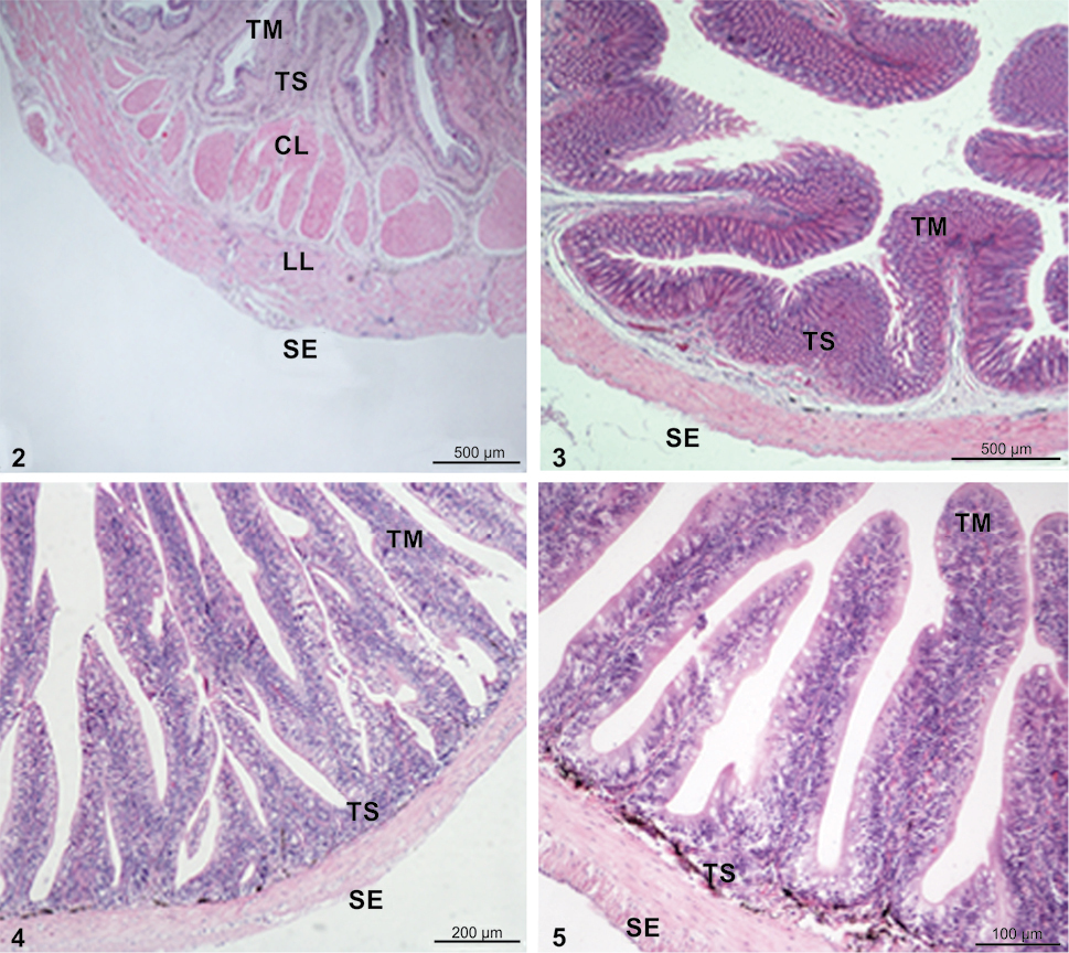
Photograph of L. crocea GIT illustrating the general organization: (2) transversal section of the oesophagus, note the muscular possessing an inner longitudinal layer and an outer circular layer; (3) transversal section of stomach showing the muscularis mucosa; (4) anterior intestine possessing longitudinal villi and thin muscular layer; (5) posterior intestine possessing short villi and thick muscular layer. Hematoxylin and eosin stain. (TM) Tunica mucosa, (TS) tunica submucosa, (CL) circular layer, (LL) longitudinal layer, (SE) serosa. |