|
||
|
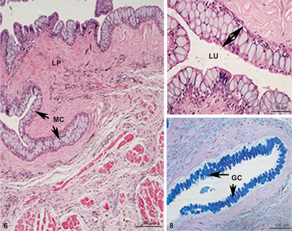
Photograph illustrating the transverse section of oesophagus containing mucus cells (arrow), lamina propria and lumen (6–7 stained with Hematoxylin and eosin stain). (8) Mucus-secreting cells in the anterior part of the oesophagus (arrows) possessing neutral and acidic carboxylated and sulphoglycoprotein. Combined stain of Periodic acid-Schiff + Alcian blue pH 2.5. (MC) Mucus cells, (LP) lamina propria, (LU) lumen, (GC) goblet cells. |