|
||
|
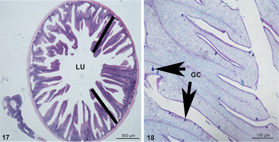
Photograph illustrating the transverse section of pyloric caeca that histologically resembles with intestine, but with exception of the higher quantity of goblet cells (arrows), (17) stained with Hematoxylin and eosin stain and (18) stained with the combined stain of Periodic acid-Schiff + Alcian blue pH 2.5. (LU) Lumen, (GC) goblet cells. |