|
||
|
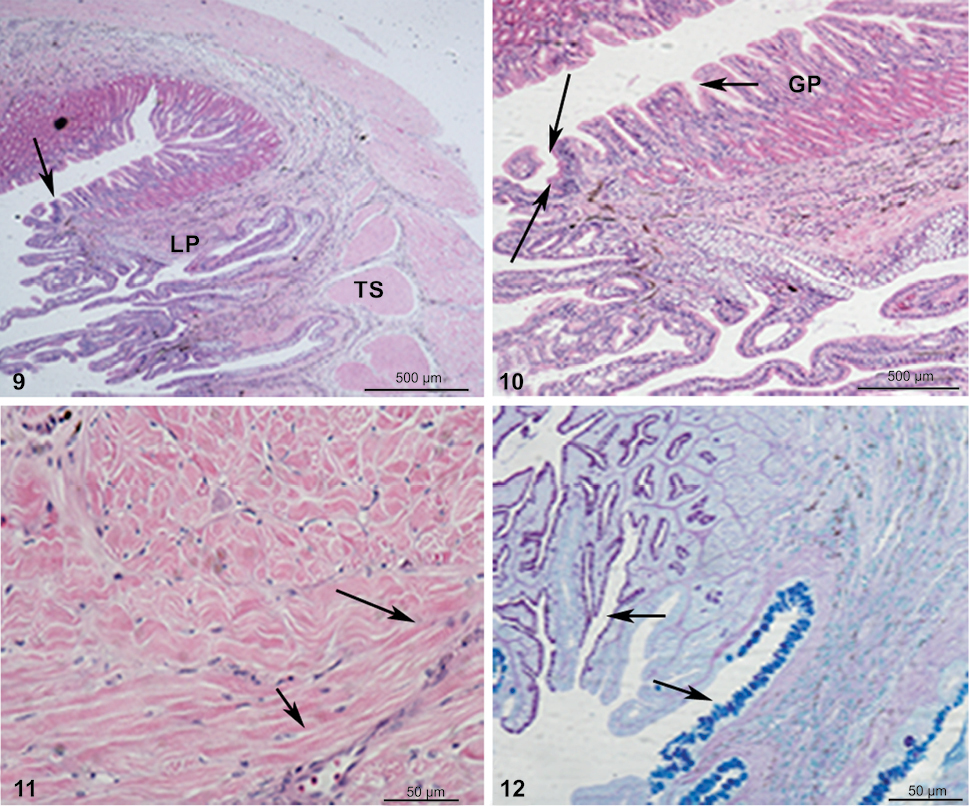
Photograph illustrating the changing of oesophageal epithelium to the gastric epithelium (arrow) in (9) and (10). Note the short non-glandular mucosal region of the gastric epithelium, (11) replacement of striated muscle by smooth muscle and (12) rapid transform of oesophageal epithelia with goblet cells (arrow) to gastric epithelia with epithelial cells containing apical mucosubstances. (9–11) stained with Hematoxylin and eosin stain and (12) stained with the combined stain of Periodic acid-Schiff + Alcian blue pH 2.5. (LP) Lamina propria, (TS) tunica submucosa, (GP) gastric pits. |