|
||
|
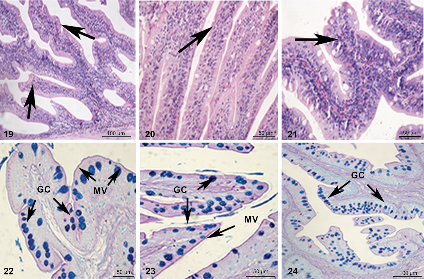
Photograph illustrating the anterior, mid and posterior regions in (19, 22), (20, 23) and (21, 24), respectively. (19–21) stained with Hematoxylin and eosin stain and (22–24) stained with the combined stain of Periodic acid-Schiff + Alcian blue pH 2.5. (GC) Goblet cells, (MV) microvilli. |