|
||
|
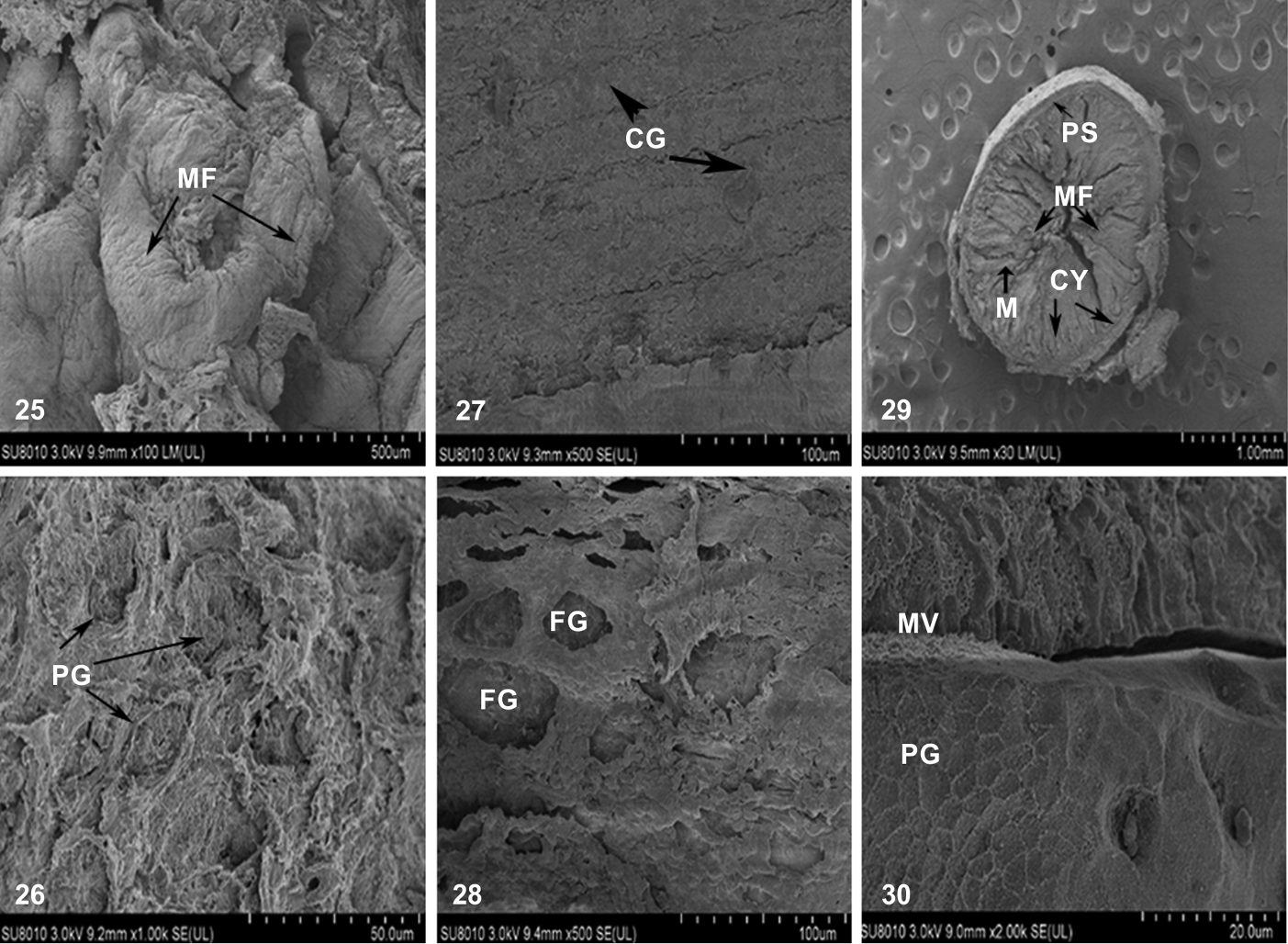
Scanning electron microscopy illustrating (25) epithelial surface of the oesophagus (26) pores of goblet cells (27) epithelial surface of the cardiac stomach showing cardiac gland (28) fundic region of the stomach showing fundic glands (29) Cross-section of the anterior intestine showing longitudinal mucosal folds, crypts, propria-submucosa and tunica muscularis (30) posterior intestine showing microvilli and pores of goblet cell. (M) Mucosa, (MF) mucosal fold, (PG) pores of goblet cells, (CG) cardiac gland, (PS) propria-submucosa, (FG) fundic gland, (CY) crypts, (MV) microvilli. |