|
||
|
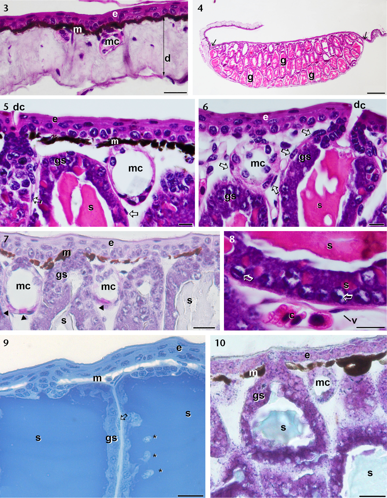
Photomicrographs of histological sections of the male inguinal gland region of O. centralis. (3–6, 8) Histological sections stained with HE. 3) Section of skin from the peripherical region of the inguinal gland. Notice that only mucous glands are present. 4) Low magnification micrograph showing the presence of many syncytial glands (g), with arrows indicating the lateral limits of the inguinal gland. (4–6) Major magnifications of the glandular apical portion, with many melanocytes (m), mucous glands (mc) and myoepithelial cells (open arrows). Note the glandular ducts (dc). 7) Histological section submitted to PAS reaction. Notice that only some cells of the mucous glands (mc) exhibit a positive reaction (arrowheads). (8) Major magnification of the lateral base portion of the syncytium, with colloidal secretion (s) in syncytium cytoplasm. Note also a blood vessel in the connective tissue. (9) Methacrylate section treated with potassium permanganate and oxalic acid and stained with Nile blue. Notice the bleached melanocytes (m) and some syncytial cytoplasmic projections (*) through the glandular secretion (s). (10) Methacrylate section stained with toluidine blue. Notice the pale blue color of the secretion suggesting it is alkaline, contrasting with the dark blue color of the glandular syncytium (gs). (e) epidermis; (d) dermis; (black open arrow) myoepithelial cells; (c) blood cells. Scale bars: 5, 6, 8 = 10 μm, 3, 7, 9, 10 = 20 μm; 4 = 200 μm. |