|
||
|
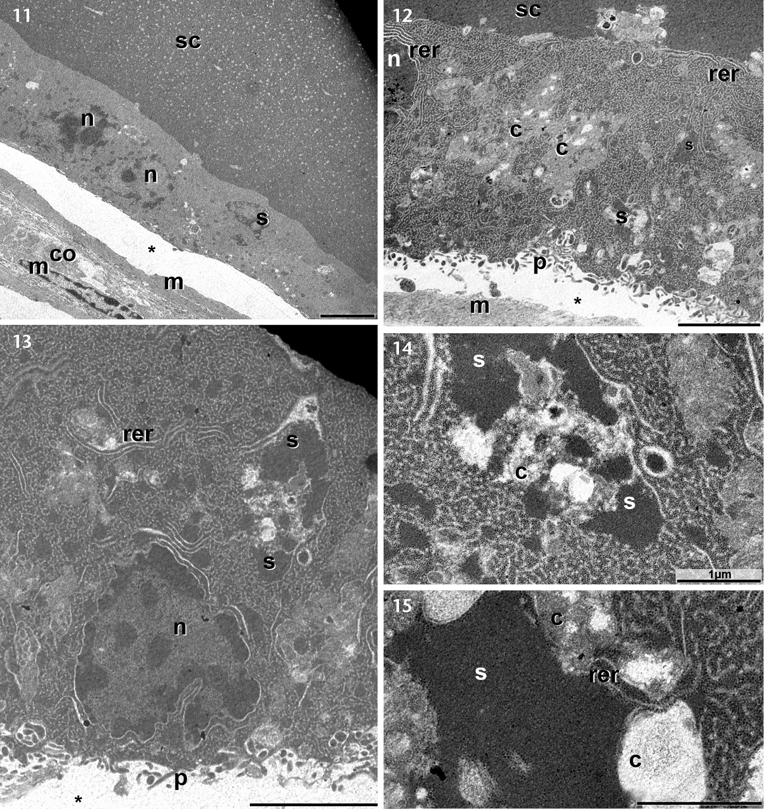
Electron micrographs of the serous glands of the inguinal region. (11) Low magnification of the secretory syncytium with two visible nuclei (n) and also a sizeable cytoplasmic secretion aggregate (s). Notice the syncytium center (sc) filled with electron dense secretion and also the clear space (*) between syncytium basis and myoepithelial cells (m). Around the myoepithelial cells are some collagen fibrils (co). (12–13) Medium magnification of syncytium, where it is possible to notice some cytoplasmic secretion aggregate (s) and some regions of the cytoplasm with medium electron density (c). (14–15) Major magnifications of two large cytoplasmic secretion aggregate, with mixed portions of electron dense secretion (s) with medium electron density cytoplasm (c). (p) basal digitiform projections; (rer) rough endoplasmic reticulum. Sacale bars: 14, 15 = 1 μm, 12, 13 = 3 μm, 11 = 5 μm. |