|
||
|
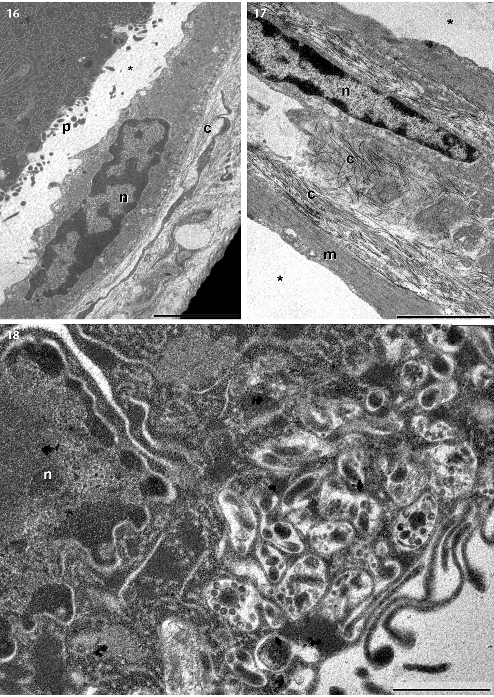
(16) The basal portion of the syncytium with digitiform projections (p) and the clear space (*) between them and the myoepithelial cells. Notice the myoepithelial cells nuclei (n) and the collagen fibrils. (17) Detail of the connective tissue between two neighbor alveoli, with myoepithelial cells (m) and collagen fibrils (c). (18) The basal portion of a syncytium with intricate projection labyrinth. Notice the syncytium nucleus with irregular outline (n). Scale bars: 18 = 1 μm, 16, 17 = 3 μm. |