|
||
|
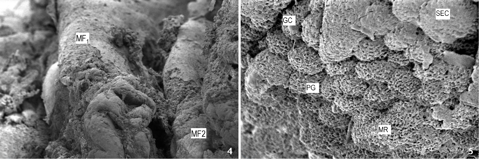
Scanning electron micrograph of esophagus showing: (4) primary longitudinal (MF1) and secondary mucosal folds (MF2); (5) stratified epithelial cells (SEC), goblet cells (GC), Pores of goblet cells (PG) and microridges (MR). Scale bar: 4 = 500 µm, 5 = 10 µm. |