|
||
|
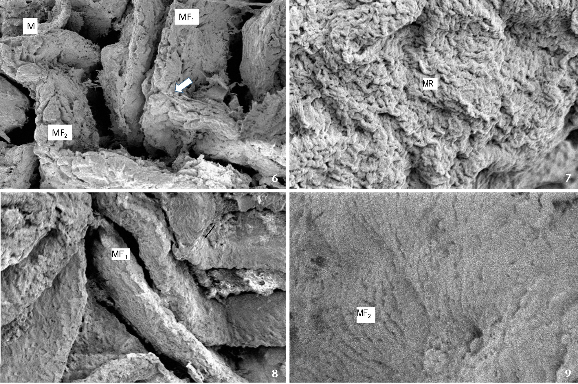
Scanning electron micrograph of intestinal bulb showing: (6) primary or major (MF1) and secondary mucosal folds (MF2), covered with thin film of Mucin (M). The zig-zag arrangement of primary folds is clearly visible in this region (←); (7) microridges (MR); (8) primary or major mucosal folds (MF1) and the zig-zag invagination (←); (9) secondary folds (MF2). Scale bar: 6 = 500 µm, 7 = 10 µm, 8 = 100 µm, 9 = 20 µm. |