|
||
|
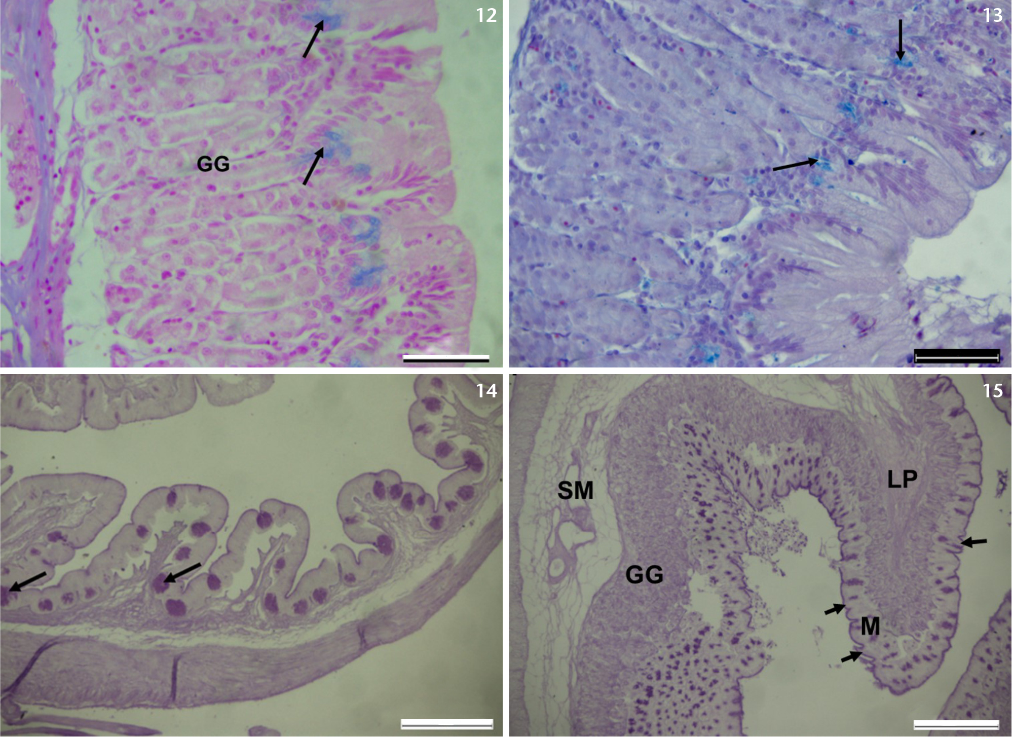
(12, 13) Photomicrographs of the cardiac stomach of T. sparrmanii showing numerous gastric glands (GG), and AB (pH 2.5) positive neck cells (arrows). (14, 15) Photomicrographs of the stomach of T. sparrmanii showing numerous gastric glands (GG), and PAS positive epithelial and mucous cells (arrows). Lamina propria (LP); Mucosa (M); submucosa (SM). Scale bars: 12–14 = 50 µm, 15 = 200 µm. |