|
||
|
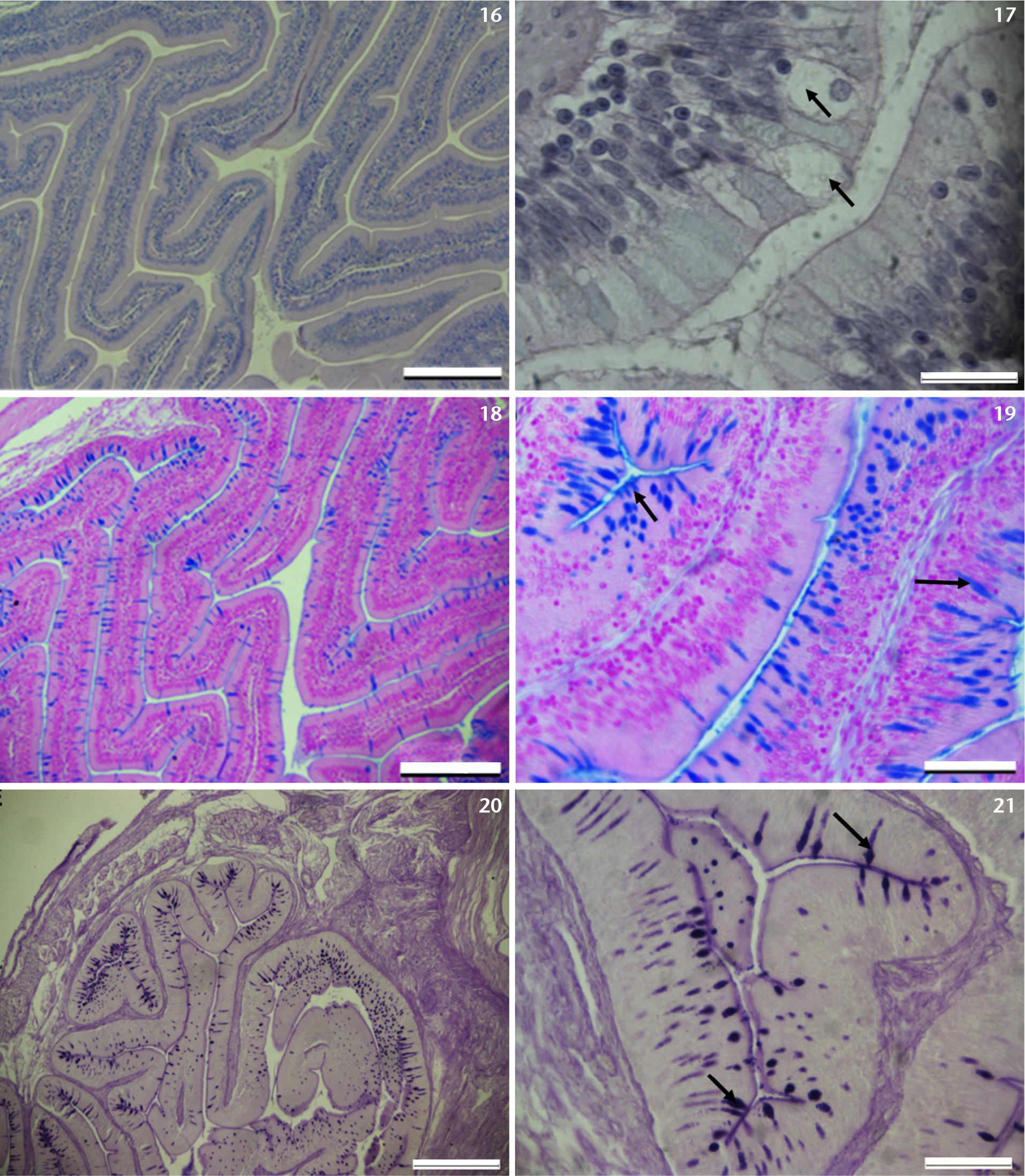
Photomicrographs of the anterior intestine (AI): (16) An overview of the anterior intestine with emphasis for zig-zag shaped villi, with columnar epithelial cells, which are well endowed with goblet cells. H&E stain. (17) Shows an enlarged image of a segment of Fig. 16 highlighting epithelial lining of the anterior intestine endowed with goblet cells (arrows). H&E stain. (18, 19) Highlights epithelial cells of the anterior intestine endowed with AB (pH 2.5) positive goblet cells. Note the elongated and oval shaped positive cells (arrows) in Fig. 19. (20, 21) Highlights epithelial cells of the anterior intestine endowed with PAS positive goblet cells. Note the teardrop shaped PAS positive cells (arrows) in Fig. 21. Scale bars: 16, 18, 20 = 200 µm, 17 = 20 µm, 19, 21 = 50 µm. |