|
||
|
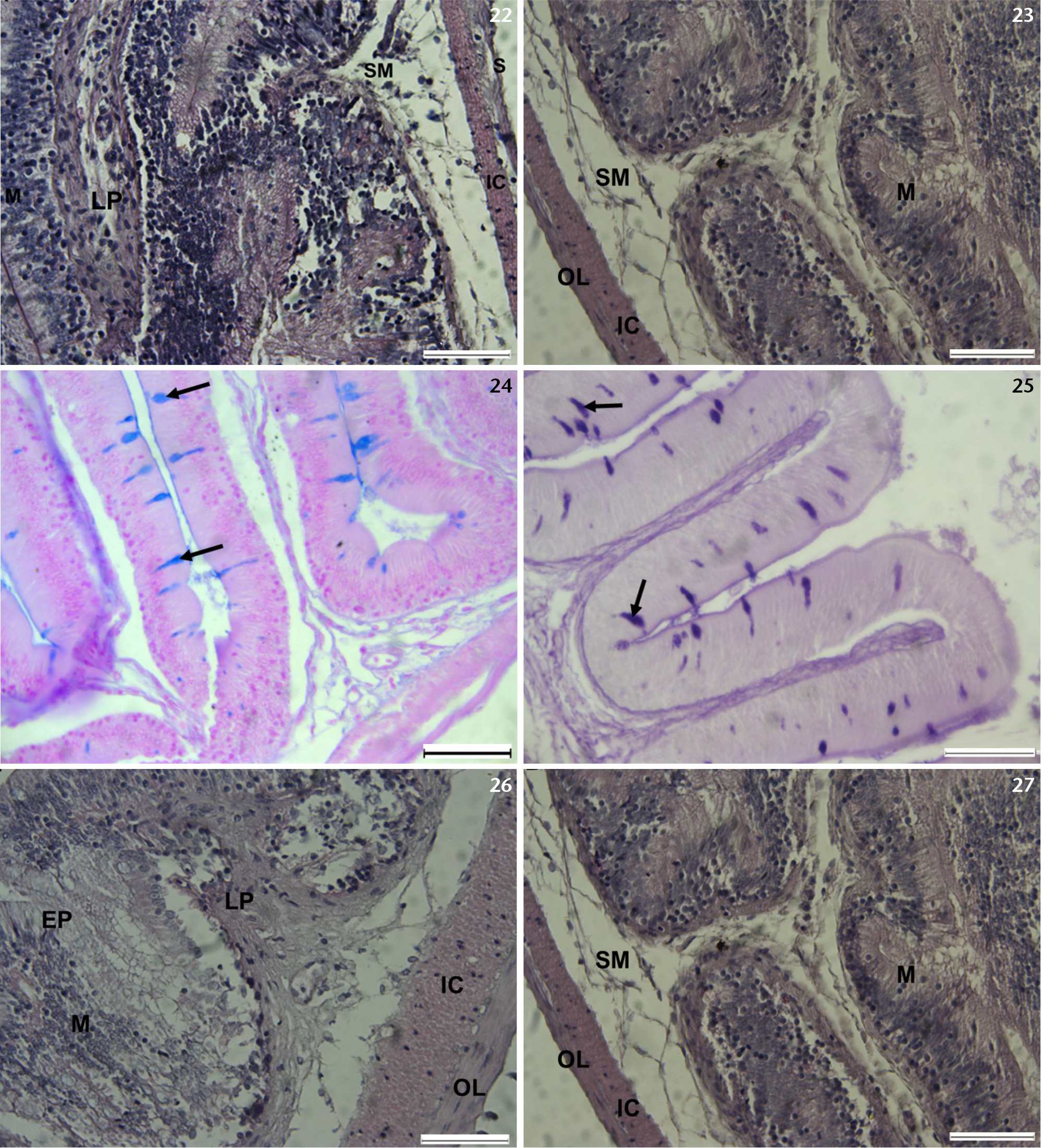
Photomicrographs of the middle and posterior intestine of T. sparrmanii. (22, 23) Photomicrographs of transverse sections of the middle intestine, showing mucosa, (M); lamina propria, (LP); submucosa, (SM); internal circular muscular layer, (IC); external longitudinal muscle layer, (OC) H&E stain. (24) Photomicrographs of transverse sections of the middle intestine, showing AB (Ph 2.5 positive cells (arrows). (25) Photomicrographs of transverse sections of the middle intestine, showing PAS positive cells (arrows). (26, 27) Photomicrographs of transverse sections of the posterior intestine, showing mucosa, (M); lamina propria, (LP); submucosa, (SM); internal circular muscular layer, (IC); external longitudinal muscle layer, (OC); serosa (S) and the epithelium (EP) H&E stain. Scale bars: 50 µm. |