|
||
|
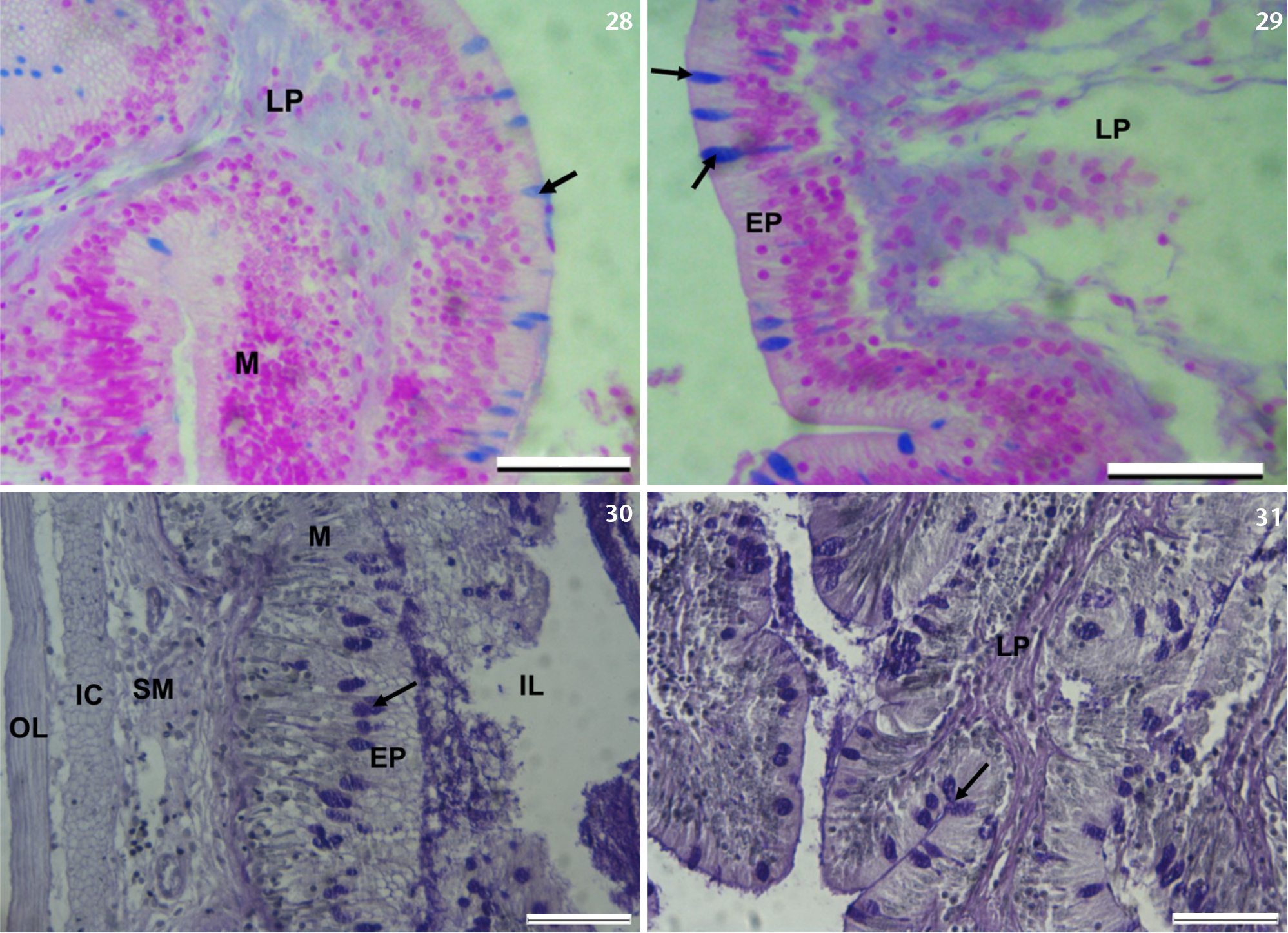
Photomicrographs of the middle and posterior intestine of T. sparrmanii. (28, 29) Photomicrographs of transverse sections of the posterior intestine, showing AB (Ph 2.5 positive cells (arrows). (LP), lamina propria; (M), mucosa; (EP), epithelium. (30, 31) Photomicrographs of transverse sections of the posterior intestine, showing PAS positive cells. PAS/haematoxylin stain. (LP), lamina propria; (M), mucosa; (EP), epithelium. |