|
||
|
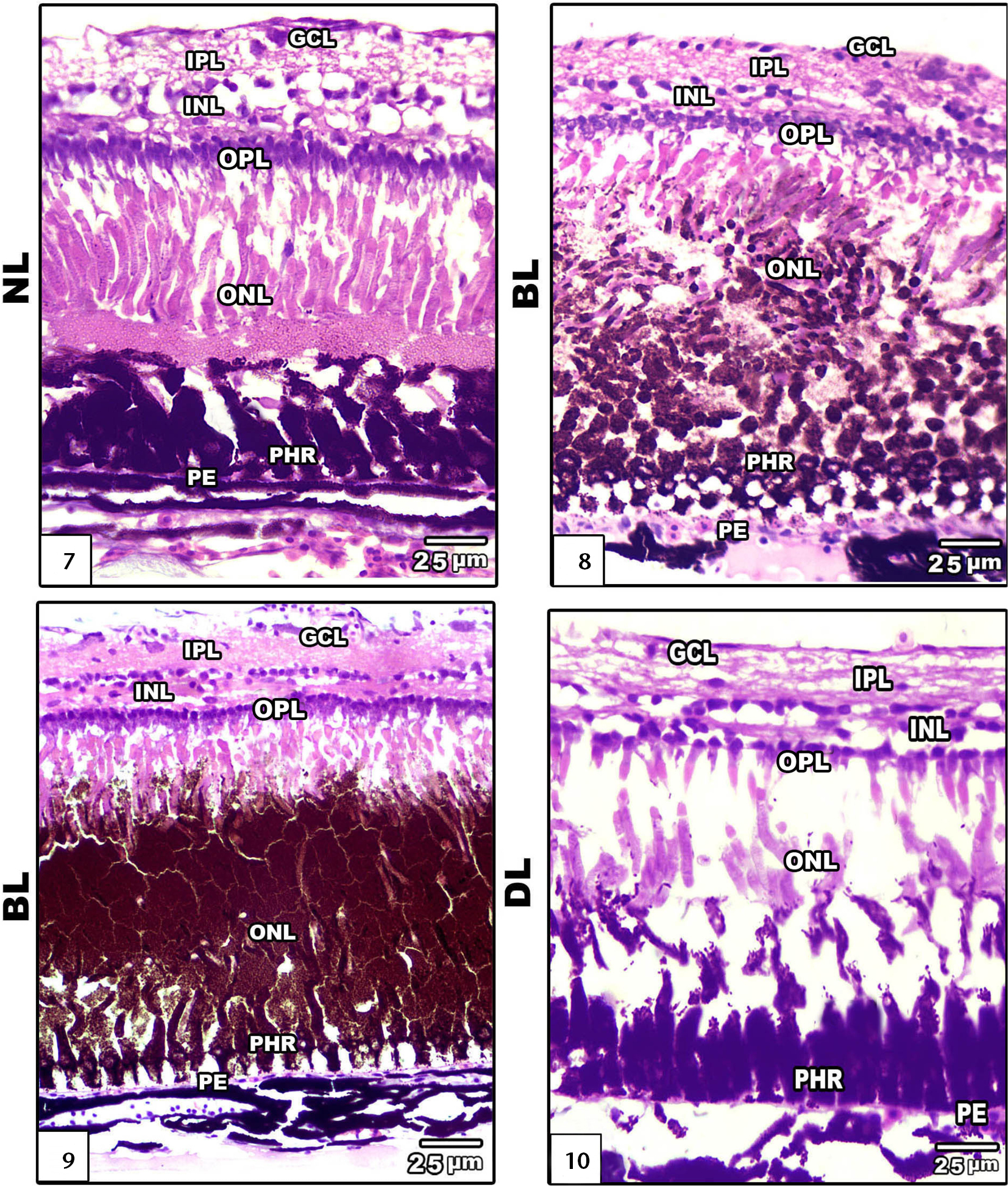
Photomicrograph of sagittal histological sections of retina of Clarias gariepinus, showing ganglion cell layer, inner and outer plexiform layer, inner and outer nuclear layer, photoreceptor layer: (7) normal light showing ordinary retinal structure; (8–9) exposure to bright light showing damaged photoreceptor and increased infiltration of dark-brown pigments; (10) dim light exposure showing regenerated photoreceptors layer and less dense nerve fibers in the outer plexiform layer and outer nuclear layers. (GCL) Ganglion cell layer, (IPL) inner plexiform layer, (INL) inner nuclear layer, (OPL) outer plexiform layer, (ONL) outer nuclear layer, (PHR) photoreceptors, (PE) pigmented epithelium, (NL) normal light, (BL) bright light, (DL) dim light. |