|
||
|
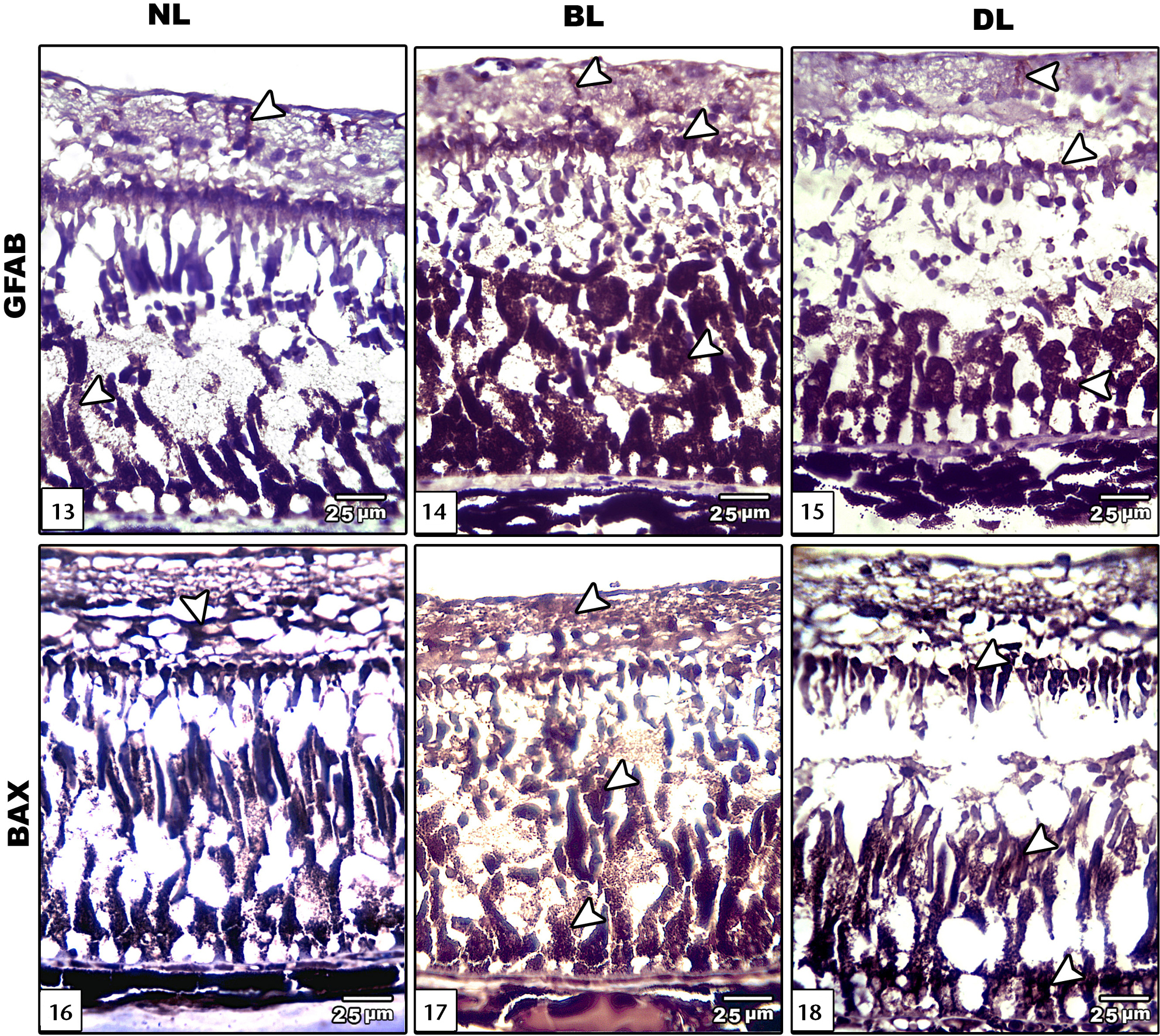
Photomicrograph of sagittal histological sections of retina of Clarias gariepinus: (13–15) showing GFAP immunostaining: (13) control showing decreased GFAP immunohistochemistry; (14)exposure to bright light showing increased immunohistochemical reaction; (15) dim light exposure showing comparatively decreased immune reaction compared to bright light;(16–18) showing BAX immunostaining. Strong reaction appeared in different retinal layers of BL and DL retina more than in normal retina. (NL) Normal light, (BL) bright light, (DL) dim light. |